| Home | AmMin | GMR | RiMG | Collectors Corner | Directory | Short Courses | |
|
|
|||||||

|
|
Volume 13, pages 297-329, 1928 MINERALOGICAL NOTES ON FRANKLIN AND STERLING HILL, NEW JERSEY CHARLES PALACHE The Harvard Mineralogical Museum has received, during the past year, very large additions to its series of specimens from Franklin Furnace, the study of which has added many new facts to our knowledge of a number of the Franklin minerals. The Stanton Collection, purchased after the death of Mr. Stanton and rich in the discoveries of the past few years, has been as yet only partly studied. The Canfield duplicates, purchased from the trustees, contained many interesting specimens. Most valuable of all; however, are the materials sent at frequent intervals by Messrs. Bauer and Jenkins, chemists of the New Jersey Zinc Company, for they are generally accompanied by careful chemical analyses and represent the newest discoveries both at Franklin and Sterling Hill. The following notes, compiled by the author whose name alone appears in the title, rest upon such analyses and upon crystallographic and optical studies made by Messrs. L. W. Lewis and H. Berman. They treat of the occurrence, crystallography, optical and chemical properties of the following minerals: azurite, bornite, cahnite, clinohedrite, clinozoisite, crocidolite, gageite, glaucochroite, hetaerolite, hodgkinsonite, leucophoenicite, manganite, quartz, smithsonite, sussexite, tephroite, tennantite, and willemite. AZURITE. While long known at Franklin as a stain associated with small veins containing chalcopyrite, azurite was but recently found in crystallized specimens. In the Stanton Collection it is represented by material from both Franklin and Sterling Hill. The Franklin crystals occur in thoroughly oxidized vein material found in the mine in the 720 pillar on 200 level. The small crystals are rich in forms. They line cavities in limonitic calcite along with malachite and fine rosettes of aurichalcite, the hydrous carbonate of zinc and copper.
The individual crystals are elongated parallel to the b axis with θ( PLATE IV |
  4a 4b EXPLANATION OF PLATE IV
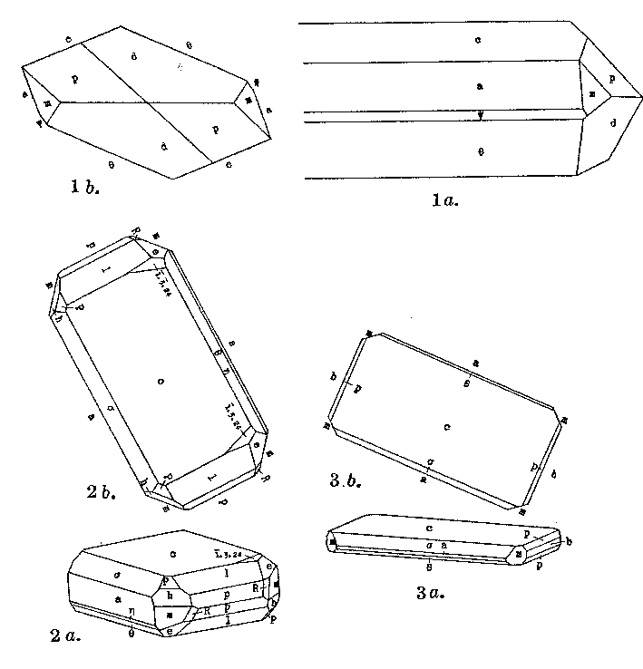
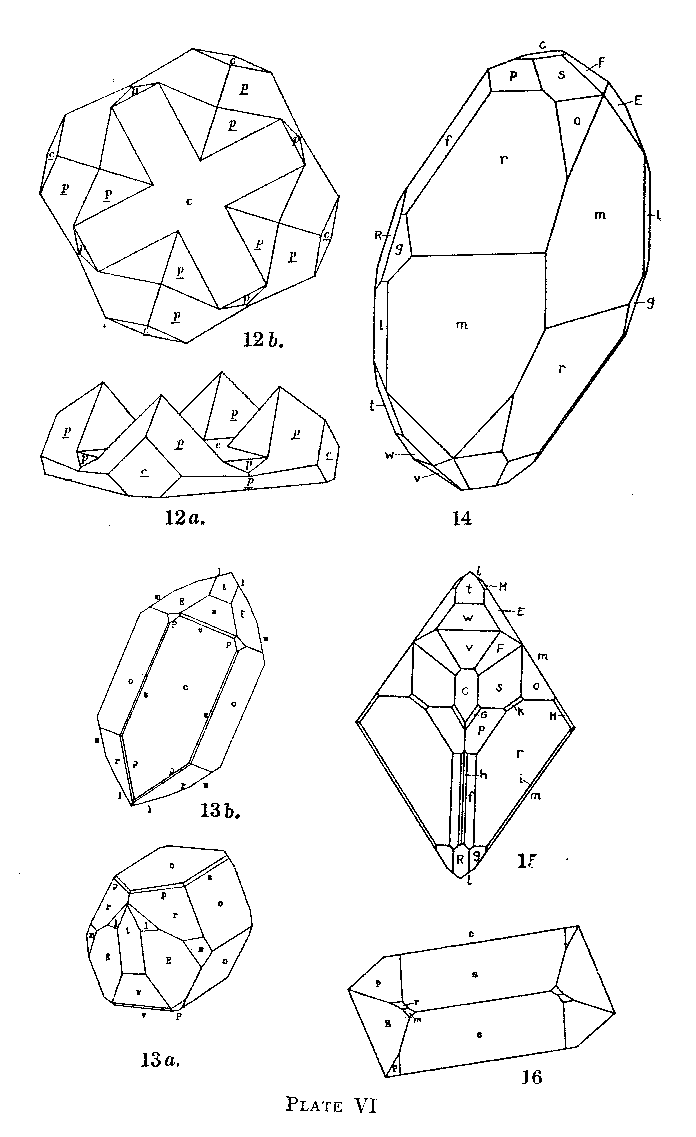
Fig. 1a. Azurite projection of a crystal showing the forms a(100), c(001), θ( Fig. lb. Azurite. The same crystal as la projected on b(010).
Figs. 2a and b. Azurite. Clinographic and basal projections of a crystal showing the forms c(001), a(100), σ(101), θ(
Figs. 3a and b. Azurite. Clinographic and basal projections of a crystal showing the forms c(001), a(100),
b(010), θ(101), σ
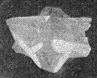
( Fig. 4a. Cahnite. Photograph of a twin crystal.
Fig. 4b. Drawing of the same crystal as shown in the photograph of 4a showing the forms a(100), m(110), p(111),
o(1
At Sterling Hill specimens of massive coarse granular franklinite ore were found intersected by veins of calcite and azurite which appear to be a replacement of the ore. As a whole, azurite followed calcite but the deposition of the two overlapped in part. The veins are generally granular but in a few cases cavities in the franklinite representing complete solution of large grains are lined with brilliant crystals of azurite. Seven crystals were measured-the largest being 2 mm. in greatest length parallel to the orthodome zone. The faces were small and signals faint but the angles check closely with those calculated from the axes based on the measurements of Tsumeb crystals.1 The crystals are flattened parallel to c(001) with the orthodome and clinodome zones prominent. Plate IV, figure 2, illustrates a small, doubly terminated crystal found with ideal development. The following forms were identified on crystals of this type: c(001), a(100), σ(101), θ( The form marked with an asterisk is an etch face occurring symmetrically developed on most of the crystals. Only on one crystal was it possible to get distinct signals and the readings were as follows:
The form is not considered of any value except as an interesting symmetrical and persistent occurrence of an etch face. Plate IV, figure 3, illustrates the habit of some paper-thin plates of azurite flattened parallel to c(001). The relative thickness is exaggerated in order to show the forms present as small faces. BORNITE. The occurrence of bornite at Franklin is uncommon although some specimens of chalcopyrite were seen which seemed to contain minute grains of the darker colored bornite. The following analysis, however, made on a specimen from the mine at Franklin by the chemists of the New Jersey Zinc Company proves its presence although mixed with sphalerite: ANALYSIS OF IMPURE BORNITE BY L. H. BAUER
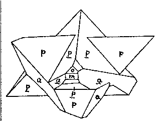
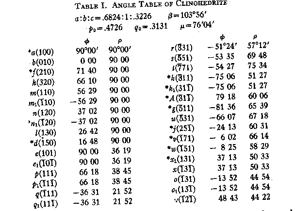
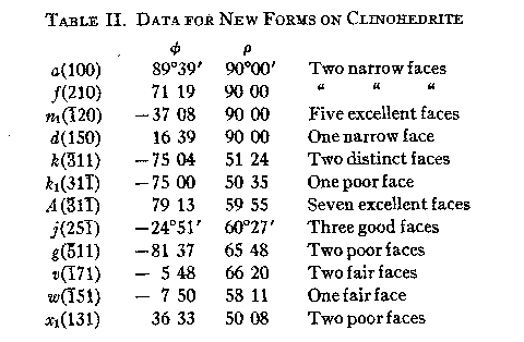
In the specimen yielding this analysis, the sphalerite was visible as abundant grains of dark brown color. It was concluded that the zinc, manganese, and a small portion of the iron were derived from the sphalerite and calculation on this basis shows 40 per cent. of the latter to be present. Deducting this and neglecting the silica as unessential the figures of the last column are obtained which yield very closely the bornite formula Cu5FeS4. CAHNITE. This mineral, first described2 a year ago in the form of crystals of minute to microscopic size, was found during the past summer in a small pocket in what may fairly be called gigantic crystals for this substance. The pocket was in 229 pillar, 20' from the 700 level north, near where Mr. Stanton first found cahnite in a rhodonite vein. The crystals were implanted on rhodonite and numbered in all about a dozen. They range in size from 2 cm. (about 3/4 inch) square, downward, are all twinned in the habit previously described for the mineral and present no new forms as they have been etched and slightly rounded. Plate IV, figures 4a and 4b, shows a typical specimen in photograph and drawing. I am indebted to Mr. Bauer for the opportunity of securing these remarkable crystals and to Mr. Lewis for photographing and measuring them. CLINOHEDRITE. Clinohedrite was first described in 1898 by Penfield and Foote3 and nothing has been added to their description of this interesting mineral in the ensuing thirty years. The material which they described came from the dump of the Parker Shaft and the mineral was not found in place at that time. The recent discovery of clinohedrite by Mr. Bauer in association with the two new lead silicates larsenite and calcium-larsenite4 seems a fitting occasion therefore to publish such new facts as have come to the writer's notice concerning it. These facts include new crystallographic data and two new analyses by Mr. Bauer, which serve to confirm the correctness of the original description, As no table of position-angles for clinohedrite has been published these values are presented in TABLE I. The table includes the forms found by Penfield and, a number since observed, marked with an asterisk, the data for which are contained in TABLE II. TABLE I. ANGLE TABLE OF CLINOHEDRITE
From time to time crystals of clinohedrite, probably from the original locality, have come into the writer's hands which yielded the combinations of forms shown in Plate V, figures 5, 6, and 7. Two other specimens in which hodgkinsonite was associated with clinohedrite contained measurable crystals of the latter which are shown in Plate V, figures 8 and 9. These are probably from a second occurrence but its location in the mine is unknown. In 1928 specimens were found in the mine at Franklin on the 400 level in the north end which contained clinohedrite associated with a number of other species including larsenite as mentioned above. The clinohedrite is fairly abundant in clusters of prismatic crystals lining cavities and intergrown with needles of larsenite. It is snow-white or glassy; transparent, with optical properties identical with those of the first described occurrence. The prismatic habit is developed parallel to the zone [101] containing the forms e, p, b and p1. These crystals are shown in Plate V, figures 10 and 11, and are markedly different in appearance from any hitherto described. All the new forms were found on the above crystals, the measurements obtained constituting TABLE II. The angles measured both on these and on the known forms conform closely to those calculated from Penfield's axes. PLATE V |
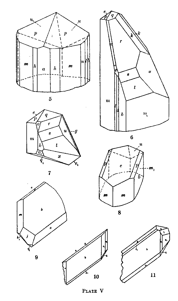
EXPLANATION OF PLATE V
Fig. 5. Clinohedrite crystal showing a(100), b(010), h(320), m(110), ml(
Fig. 6. Clinohedrite. Projection on b(010) of a crystal showing b(010), m(110), ml(
Fig. 7. Clinohedrite. Projection on b(010) of a doubly terminated crystal showing b(010), m(110), e(101),
el(
Fig. 8. Clinohedrite crystal showing b(010), m(110), ml(
Fig. 9. Clinohedrite. Projection on b(010) of a crystal showing b(010), m(110), e(101), p(111),
ql(11
Fig. 10. Clinohedrite. Projection on b(010) of a crystal showing b(010), ml(
Fig. 11. Clinohedrite. Projection on b(010) of a crystal showing b(010), ml(
TABLE II. DATA FOR NEW FORMS ON CLINOHEDRITE
CHEMICAL ANALYSES OF CLINOHEDRITE
1. Analysis by Foote, 1898, average of two, Parker Shaft.
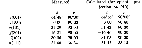
Mr. Bauer analyzed a small sample (.183 gram) of the newfound clinohedrite which had been purified as completely as possible by Mr. Berman. It was impossible to free it entirely from larsenite and calcite. It is shown together with a second analysis of material from the Parker Shaft in the table below. They are essentially identical and yield the original formula, H2CaZnSiO5. The rediscovery of clinohedrite in the mine at Franklin leaves only one of the numerous species described by Penfield from the Parker Shaft dump to be found again, namely nasonite. Roeblingite occurs in small amount with the new find of clinohedrite. CLINOZOISITE. This mineral, not before described from Franklin, was identified by crystal form and optical characters among the many minerals coating the walls of the cavities from which gageite was first described. The principal wall coating is orange colored zincite upon which rest pyrochroite, willemite needles, gageite and calcite. The clinozoisite is in flattened prisms, deeply striated in the direction of their length and terminated by a single pair of pyramid faces with the angles and in the familiar habit of epidote. They are white to pale yellow in color and show basal cleavage. Three crystals were measured, yielding poor reflections but sufficient to identify their forms according to the following table of angles.
Since the angles of epidote and clinozoite are practically identical, the identification of these crystals as belonging to the latter species rests on their optical properties which were determined by Mr. Berman as follows: Biaxial negative. Extinction parallel on length sections. Z ^ c = 31°. Y=B, X=a. Dispersion strong, ρ>v, α=1.684; β=1.691; γ=1.698. These characters show the mineral to be closely related to clinozoisite but the indices are lower than those of any member of the zoisite group, even the newly described pumpellyite. As there is no possibility of obtaining sufficient amounts of the mineral for chemical analysis, its true place in the series must remain uncertain. CROCIDOLITE. Crocidolite has long been known from Franklin in inconspicuous narrow calcite veins in which it was associated with granular sphalerite. Recently it was found in abundance and the Stanton Collection contained numerous specimens showing its association. It is a very pale blue variety in excessively fine needles more or less felted together or contained as inclusions in other minerals. It appears to have formed vein-like masses in which the chief mineral is calcite in perfectly colorless anhedra, the exterior rounded and coated with crocidolite. Willemite is also found in similar rounded crystals of considerable size, pale green or perfectly colorless, but sometimes blue from included needles of crocidolite. Sphalerite too is found in these veins in paly green to white cleavage masses. The occurrence of needle-like crystals of quartz in one specimen is described on a later page of this paper. A sample of crocidolite was analyzed by Mr. Bauer, as shown below in Column 1. The sample showed the presence of willemite by its fluorescence so the ZnO was deducted as Zn2SiO4 willemite (8.91 per cent.) and the analysis recalculated to 100 per cent. The specific gravity of this sample is 3.195.
The formula may then be written, ignoring insignificant amounts of CaO and A1 2O3 as: 4H2O·2Na2O·6(Mg,Fe)O·3Fe2O3·20SiO2.Dr. M. A. Peacock, who has recently studied the chemical composition of the South African crocidolites 5 has kindly discussed this analysis along similar lines and reports as follows: "Crocidolite may be regarded as containing the following molecules in varying proportions:l (Fe,Al) 2O3·H2O·SiO2 (Penfield Molecule),m (Fe,Al) 2O3·H2O·4SiO2 (Gordon's ideal riebeckite molecule),n (H 2,Na2,Ca,Mg,Fe)O·SiO2 (Metasilicate amphibole molecule).In the Franklin crocidolite l is small and m and n about equal in amount. It differs from South African crocidolite in having MgO greatly preponderating over FeO instead of the reverse, and in having a greater relative amount of Fe 2O3"GAGEITE. The existence of gageite as a definite mineral species rests upon an analysis made upon a very minute amount of material (.04 gr.). In a recent paper, 6 Gordon has tried indeed to show that gageite was probably identical with tephroite. The following facts regarding this mineral seem, however, to establish its validity as an independent species.The optical characters determined by Larsen on type material have been found to be characteristic of numerous specimens of the mineral leaving no ambiguity in its determination. In order better to establish its chemical nature, a gram sample of gageite was purified by Mr. Berman from a nearly homogeneous specimen obtained by the kindness of Captain George Rowe of Franklin and this was analyzed by Mr. Bauer.
1. Analysis of gageite by.L. H. Bauer
The composition is expressed by the formula 8(Mn,Mg,Zn)O·3SiO 2·3H2O. Specific gravity 3.584 (Pycnometer, Berman).These results agree substantially with those originally obtained by Phillips, except for a somewhat larger content of water, and lead to the same formula as proposed by that author. In the Stanton Collection, there are many specimens in which gageite is associated with chlorophoenicite. No crystals were measurable. The gageite here as usual is in fibrous aggregates of pale pinkish tint and seems to be a replacement or pseudomorph after an unidentified mineral. A comparison of the chemical and optical characters of gageite with those of tephroite shows a wide divergence of nature. It would have to be compared rather to a "hydrotephroite" which is a vague name attached to various uncertain and indefinite alteration products of tephroite. There seems to the writer no good reason for denying the right of gageite to a definite place among the minerals. GLAUCOCHROITE. This mineral was established by Penfield and Warren in 1899, on material found during the sinking of the Parker Shaft and has not since been identified at Franklin. The discovery and analysis of new material of this mineral is, therefore, a welcome contribution to our knowledge of it. Mr. Bauer has sent to the writer several specimens of bluish glaucochroite in massive form intermixed with willemite, hardystonite, tephroite and franklinite which were found on the picking table. Their place in the mine is unknown except that they came from a deep level. Some of this material was purified. by Berman and gave the following optical properties. Biaxial negative. 2V medium large. α=1.68, β=1.71, γ=1.725. This sample was analyzed by Mr. Bauer as shown below. The powder gave under the iron spark-gap the willemite phosphorescence from isolated grains. The zinc found in the analysis was therefore calculated as willemite and deducted. The analysis is practically identical with the original analysis by Warren.
1. Analysis of glaucochroite by L. H. Bauer.
HETAEROLITE. This mineral was originally found in abundance at Sterling Hill in oxidized material and was first described by Moore in 1877, somewhat doubtfully as a zinc hausmannite. No further examination of the mineral was made until 1910 when the writer published a new analysis by Schaller and concluded that it was zinc hausmannite but in so doing ignored a considerable content of water which was not accounted for. In 1913, Ford and Bradley described hetaerolite from Leadville, Colorado, where also it was found amidst oxidized ores. They concluded that it should be regarded as a hydrous oxide with the formula 2ZnO·2Mn 2O3·H2O. But their material was impure and to obtain this result 10 per cent. of calamine had to be deducted from the analysis.Mr. Bauer has now supplied analyses of a new occurrence of the mineral at Sterling Hill in which it is associated with unoxidized franklinite. These show scarcely a trace of water and yield an ideal ratio for zinc hausmannite, ZnO·Mn 2O3. This material is black and crystalline and identical with crystals from Franklin, long since measured by the author, which showed form and twinning similar to but not identical with those of hausmannite. Mr. Berman has compared the optical properties of the analyzed mineral with those of the measured crystals and of the type, massive mineral and finds them all so nearly alike that there now seems no reason to doubt that hetaerolite is a zinc hausmannite.The following data establish the characters of unaltered hetaerolite. For the partly hydrated mineral hitherto described, the name hydrohetaerolite may well be employed. The crystals: from Franklin were first brought to the Harvard Mineralogical Laboratory by Mr. Cahn in 1914. They are minute black octahedroids lining druses in thin veins in massive ore, associated with jeffersonite, wine colored and pink hodgkinsonite and green willemite. Calcite sometimes fills the center of the vein. The crystals show only the unit pyramid (111) and base (001), the latter sometimes large. A few of them show the twinning illustrated in Plate VI, figures 12a and b, with four individuals in twin relation to a fifth on e(101). There is, however, generally wide departure from the ideal symmetry here shown; some individuals may be smaller than others or quite lacking. The crystals, though brilliant, are somewhat facetted so that measurements were not satisfactory. On one simple crystal, however, four identical readings were obtained for the inclination of the pyramid to the base.
The crystals from Sterling Hill are unmodified pyramids implanted on massive franklinite and show no twinning. The color of hetaerolite is shining black with a dark brown streak. The table gives in comparative form the optical characters, observed specific gravity and calculated specific gravity (Gladstone and Dale) for the minerals of this group. The last named values are a measure of the effect produced by the introduction of water in the hetaerolite molecule. TABLE I
The analysis by L. H. Bauer of a sample of hetaerolite from Sterling Hill, purified by Berman (Sp. gr. 4.85) is as follows:
This analysis is that of an anhydrous oxide and it yields the ratio demanded by the formula ZnO·Mn2O3 with great exactness. Hetaerolite is unaltered in the blowpipe and yields a zinc coating when reduced with soda on charcoal. HODGKINSONITE. Hodgkinsonite has been found in recent years in a great variety of associations at Franklin and must be ranked as one of the most characteristic members of the mineral assemblage filling the "pneumotectic veins." Mr. Bauer has made two analyses which confirm the original chemical interpretation of this mineral. Its optical orientation has been more exactly fixed by Mr. Berman's studies. And Berman and Lewis have measured a number of crystals found among the abundant material of the Stanton Collection which considerably enlarges the crystallographic interest of the species. These crystals, instead of presenting a prismatic habit parallel to the c axis which has been hitherto the rule, are dominantly developed parallel to the a axis. Some are tabular parallel to the base (001) with strong development of the clinodome zone; others have neither base nor prisms except as line faces and the clinodomes determine the prismatic habit. In addition to these novel habits these crystals have yielded some 17 new forms additional to the 24 already established, making the form series a highly complex one. The optical characters as revised by Berman are as follows:
Biaxial negative, 2V=50° to 60°
PLATE VI EXPLANATION OF PLATE VI Figs. 12a and b. Hetaerolite. Plan and clinographic projection of a twin group of five crystals showing the forms c(001) and p(111), four of the individuals twinned to the fifth one on faces of the second order pyramid.
Figs. 13a and b. Hodgkinsonite. Clinographic and basal projections of a crystal showing the forms c(001), m(110),
l(210), s(011), o(021), v(
Fig. 14. .Hodgkinsonite. Clinographic drawing with the axis b to the front, showing the forms c(001), m(110),
1(210), s(011), o(021), v(
Fig. 15. Hodgkinsonite. Basal projection of a crystal showing the forms of figure 14 with the addition of the forms h(623),
i(441), k(243), G(223), H(131), M(
Fig. 16. Hodgkinsonite. A crystal projected on b(010), showing the forms c(001), m(110), s(011),
p(111), P(
In part of the vein yielding the willemite specimens described in a later part of this paper, the willemite crystals
grow fewer or are wholly lacking. Their place is taken by scattered, implanted crystals of hodgkinsonite; bluish green
tephroite crystals with tips blackened by oxide of manganese; and white barite crystals or platy masses, which, with
calcite, sometimes wholly filled the fissure. The barite crystals and those of tephroite are of common habits but the
hodgkinsonite presents a combination of forms wholly flew to this mineral. The crystals are in part clear pink, in part
of a fine topaz yellow color and do not exceed 5 mm. in diameter. Figure 13 shows the habit of the crystals; in contrast
to all previous finds of hodgkinsonite the prism is almost lacking and the clinodome zone is dominant with o (021) and
c
(001) the principal faces.'The pyramids r (221) and E (
m(110) p(111) *U(320) P( The two forms marked with an asterisk are new and are established by the following measured angles:
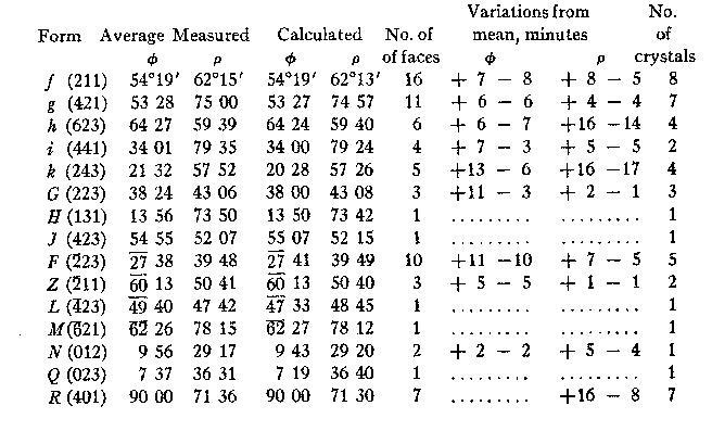
Among the specimens from the Stanton Collection, one hodgkinsonite specimen is remarkable for excellent crystals highly facetted with new forms. Figure 14, Plate VI, illustrates a find of small gem-like doubly terminated pink crystals delicately attached to larger singly terminated crystals of the same habit. One end of a doubly terminated crystal illustrated in plan by figure 15, Plate VI, measuring only 2 mm, in its greatest diameter gave reflections on one end from 44 faces of 26 forms of which 15 belonged to forms new to the mineral. The crystals are elongated parallel to the c axis with m (110) and r (221) dominant and in approximately equal development. Eight excellent crystals were measured and the new forms are well established. The following table summarized the observations establishing the new forms:
The other forms present and shown in the drawing are c(001), m(110), 1(210), s(011), o(021),
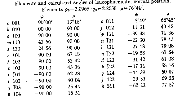
v( LEUCOPHOENICITE. Little of importance has been added to the physical and chemical data relating to leucophoenicite since it was first described by Penfield and Warren8 excepting the optical constants contained in Larsen's Tables and the crystal elements and forms published by the author,9 and reproduced in Goldschmidt's Atlas der Krystallformen. The angle tables and drawings as here given were already prepared in 1910 but are now first published.Leucophoenicite. Axial ratio: a: b : c=1.1045:1: 2.3155:β=76°44'.
HABIT. Commonly in isolated crystalline grains or massive granular. Crystals rare, generally of epidote habit, more or less elongated parallel to the b axis, with striated orthodome zone; but also tabular parallel to the orthopinacoid or an orthodome. Crystals generally minute and frequently twinned on the base, either in contact or interpenetrating and with numerous lamellae in parallel growth. The crystallization of leucophoenicite is here first described in detail, Penfield, who established the species, being unable to determine on his material even the crystal system. The first crystals measured by the writer came from a specimen in the collection of Mr. Canfield who most kindly consented to sacrifice a part of it for the purpose. Some ten crystals, combinations numbers 1 to 8, and figures 17 and 18, Plate VII, proved measurable but many were fragmentary and none were of first class quality. The measurements were made on the two-circle goniometer, the crystals, owing to their peculiar habit, being mounted so that the orthodome zone was vertical with (010) as pole. The measurements and calculated angles together with the elements and symbols of the forms in this position are contained in TABLE I. | |||||||||||||||||||||||||||||||||||||||||||||||||||||||||||||||||||||||||||||||||||||||||||||||||||||||||||||||||||||||||||||||||||||||||||||||||||||||||||||||||||||||||||||||||||||||||||||||||||||||||||||||||||||||||||||||||||||||||||||||||||||||||||||||||||||||||||||||||||
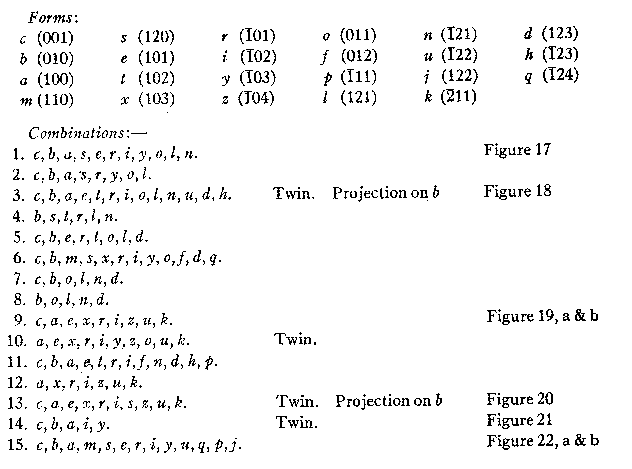
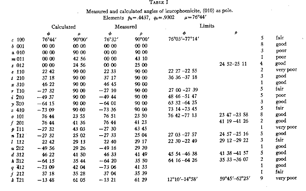
In TABLE II may be found the elements and symbols transformed to the normal position with the calculated angles for the forms. The letters and order of listing serve to identify the corresponding forms in the two tables. The calculation of the elements is based on the measurements given by 27 faces of 6 forms on 8 crystals, only those faces which gave sharp images on the goniometer being employed. The position chosen was that which gave the simplest symbols and at the same time brought the twin and cleavage plane into the position of basal pinacoid. No interpretation of the highly peculiar assemblage of forms offered the slightest resemblance to the form series of any member of the humite group to which leucophoenicite is related chemically. TABLE II
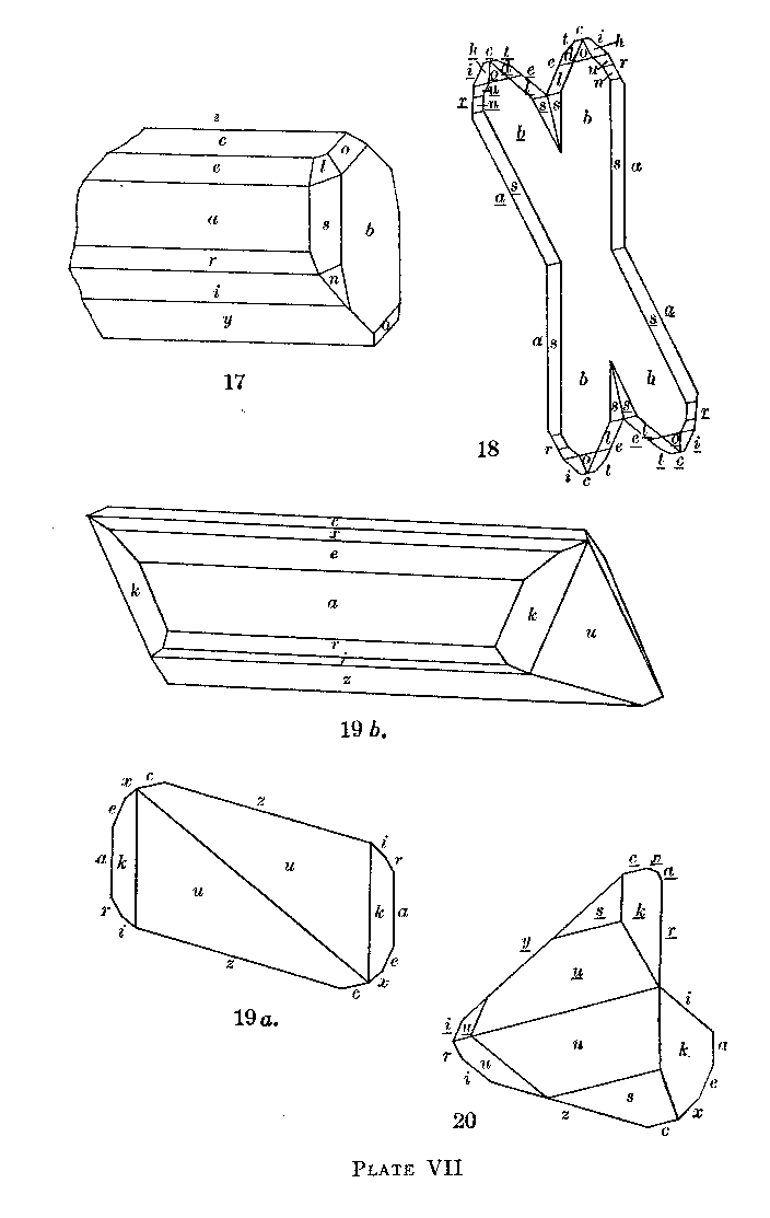
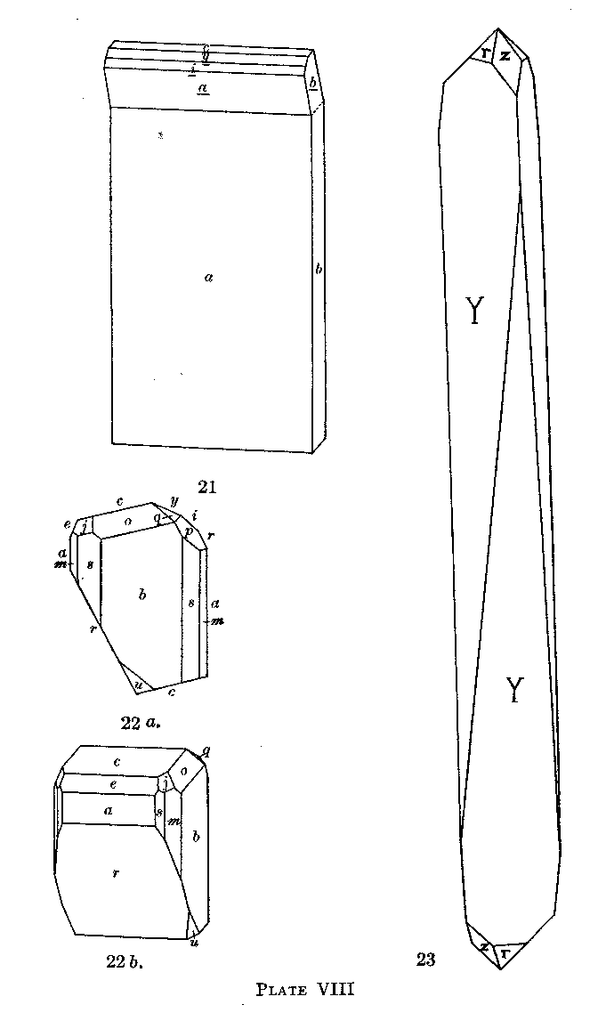
Much has been learned since its original description of the occurrence of leucophoenicite in the mine at Franklin. It is now known to be a very common mineral there, constituting veins and replacement areas in the ore and has frequently been found in place. Two occurrences especially were observed and described to the author by Mr. Hodgkinson, formerly of the Mine staff at Franklin. One consists of a single small cavity in ore, an opening in a vein consisting of leucophoenicite and willemite in granular admixture. This cavity is lined with pale yellow drusy garnet upon the surface of which are implanted crystals of leucophoenicite and willemite and a single white calcite scalenohedron. The willemite is pale green, long prismatic in habit and brilliant in lustre. The leupophoenicite is clear red in color in striking contrast to its background. The crystals are slender prismatic forms both simple and twinned, the longest doubly terminated, about one third of an inch in length and showing the forms of Plate VII, figure 19. The twin shown in Plate VII, figure 20, and the combinations 10, 11, and 12 are from this specimen. These crystals have suffered somewhat from etching so that the measurements given were poor but they sufficed to establish the forms, characteristic for which are the orthodome z and the pyramids u and k. Another occurrence of leucophoenicite was found by Mr. Hodgkinson in place in the northern end of the mine near the hanging wall of the west leg of the ore body, within two feet of a dike of pegmatite. It occurs in a continuous seam with swells and pinches, the former making vugs in which the crystals have formed. The specimens at hand show walls of banded ore with much franklinite which on the boundary of the cavity is in isolated cubic crystals. The walls of the central cavity are coated with gray calcite, merging inward to pale rhodochrosite, poorly crystallized in parallel groups of rhombohedra. Upon the carbonates is a coating of silky, felted sussexite, often in a thin film adhering closely. A central mass four inches or more across consists of massive, dull-brown leucophoenicite, becoming crystallized towards the center, either in the slender plate-like crystals shown in Plate VIII, figure 21, the broad surface deeply striated by twinning, with bright faces of the base or basal cleavage; or in rarer, isolated, stouter, more brilliant crystals like Plate VIII, figure 22. These are clear pink and of a lively color, the plates dull brown, clear to opaque. In some cases groups of the platy crystals are aggregated in fan-shaped groups, rising from the massive matrix. These specimens have added much to our knowledge of the crystallography of leucophoenicite and are much the most attractive yet found of this peculiar mineral.
PLATE VII
EXPLANATION OF PLATE VII
Fig. 17. Leucophoenicite. Crystal showing c(001), b(010), a(100), s(120), e(101), r(
Fig. 18. Leucophoenicite. Projection on b of interpenetrating twin crystals twinned on c(001). A combination of c(001),
b(010), a(100), e(101), t(102), r(
Fig. 19a. Leucophoenicite. Projection on b of a crystal showing c(001), a(100), e(101), x(103),
z( Fig. 19b. Leucophoenicite. The same crystal as shown in 19a in clinographic projection showing the prismatic development parallel to the b axis.
Fig. 20. Leucophoenicite. Projection on b of a crystal twinned on c(001), showing c(001), a(100),
s(120), e(101), x(103), r(
MANGANITE. The monohydrate of manganese has not previously been reported from Franklin. A specimen which proves to contain this mineral has been received from Mr. Bauer, It consists of a small fragment of compact limestone with a network of vugs lined with calcite crystals, some of which are very brilliant. Implanted on these are clusters of slender radiating needles, black and lustrous, which by chemical tests and crystal form proved to be manganite. The specimen was found in the mine at Sterling Hill but the exact locality is not given. Although the crystals are bright they are much striated and yielded poor measurements. These, however, sufficed to show the presence of the forms x(100), k(230) and u(101). In another specimen loaned by the U. S. National Museum the manganite crystals line cavities in a massive yellow andradite garnet.
QUARTZ. Quartz has until recently been a mineral rather conspicuous at Franklin by its absence. Numerous specimens in
the Stanton Collection, however, show it in veins in massive ore with wall surfaces coated with drusy crystals. And it
is also found in cavities in carbonate veins in crystals of ordinary habit. It is, however, to call attention to a very
peculiar specimen from Franklin presented to the Museum by Mr. Bauer that the mineral is here mentioned. This is a vein
in ore, the walls of which are lined with rhombohedral calcite crystals on which are minute plates of hematite. The
whole cavity of the vein is filled with a felted mass of the finest crocidolite fibre, pale blue in color; lying loose
in the felt or slightly attached to the wall by one end are needles of quartz, colored faintly blue by inclusions of
crocidolite. These needles range from minute spicules to slender rods 3 cm. long and 3 mm. in diameter. They are of
trigonal cross section and represent extremely steep rhombobedrons, doubly terminated and without visible planes of the
prism, much more like calcite forms than any quartz crystals known to the writer. The tip of the crystal may be
needle-sharp; or it may be terminated by the faces of the positive and negative unit rhombohedrons. Although there are
no visible prism faces and the crystal planes are dull, faint reflections were observed at 90°, the prism position,
doubtless caused by minute striations. Readings were obtained from the rhombohedron faces only by wetting with alcohol
or attaching glass slips with a film of liquid. The average reading obtained was ρ=87°30' which agrees most nearly with
the form Y(18.0.
PLATE VIII
EXPLANATION OF PLATE VIII
Fig. 21. Leucophoenicite. Unsymmetrical twin on c(001). Showing on one half a(100), and b(010), and on the other half
a(100), b(010), c(001), i(
Fig. 22a and b. Leucophoenicite. Projection on b and clinographic projection of a crystal showing c(001),
b(010), a(100), m(110), s(120), e(101), r(
Fig. 23. Quartz. Drawing of a crystal showing the rhombohedrons r(10 The only comparable quartz crystals which have been figured are shown by LaCroix10 from La Gardette.
SMITHSONITE. The carbonate of zinc has long been known both at Franklin and Sterling Hill in massive, earthy, or
spheroidal forms. Measurable crystals, always rare in smithsonite, have not been found there until recently upon a
specimen saved on the picking table at Franklin. Mr. Bauer sent this specimen to the Harvard Museum for determination.
It is a fragment of coarse franklinite-willemite ore with two or three solution cavities some lined with needles of
calamine and one with pure white crystals of smithsonite. The crystals are of typical calcite habit, the scalenohedron v(21
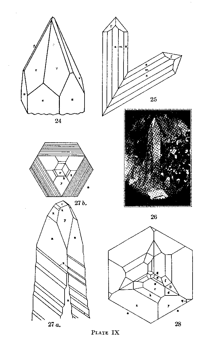
Figure 24, Plate IX, shows the habit of the crystals. So far as can be found by examination of the literature no scalenohedron has been measured or figured for smithsonite; while this form v is listed it must have been determined only by inspection. The material at hand is insufficient for analysis but its quality indicated great purity and invited optical study. Mr. Berman measured the indices and obtained results presented together with comparative data in the following table. SMITHSONITE - OPTICAL DATA (Immersion media used for determinations)
1. Not analyzed. Crystal data above. Optical properties indicate that material is quite pure. The foregoing table indicates that the optical properties of smithsonite are fairly constant for various compositions within a short range. Although the few figures given in textbooks and reference books on mineralogy vary markedly from the above given data, there seems to be no reason to suppose that such a variation exists. The highest value given for the refractive index in the calcite group is that for siderite, ω =1.875 for pure FeCO3; it seems, therefore, unlikely that the smithsonite from Moresnet, Belgium, should have for its index of refraction 1.872 as reported by Gaubert.11 The optical constants given by Mountain, number 5 of the table, are probably very close to those for pure ZnCO3. SUSSEXITE. This borate of manganese and magnesium is one of the minerals peculiar to Franklin. It has been found in a variety of associations at different times but never in large amounts. Recently there was found on the picking table a specimen so different in appearance from the normal sussexite that Mr. Bauer made an analysis which, with a specimen of the mineral, he has kindly placed at my disposition. It shows a narrow vein in massive ore largely composed of yellowish willemite with which is intermingled a dull pink massive substance looking somewhat. like garnet, and some carbonate. The pink mineral is the sussexite; when crushed it shows under the microscope a felted fibrous structure quite unlike the ordinary parallel and separable-fibrous texture of sussexite. The optical data, however, and the analysis show it to be that mineral. In it, however, there is much less magnesium than in the type material and correspondingly more manganese. It is, therefore, quite a distinct facies of sussexite. The optical characters were determined by Berman as follows:
Biaxial negative. 2V small, elongation negative, parallel extinction. α=1.65; β=1.71; γ=1.715.
PLATE IX
EXPLANATION OF PLATE IX
Fig. 24. Smithsonite. Drawing of a crystal showing the forms m(10 Fig. 25. Tephroite. Projection on a(100) of a twin crystal, twinned on k(011), showing the forms a(100), b(010), m(110), s(120), and f(121).
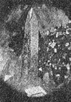
Fig. 26. Willemite. Photograph of the crystal shown in the next figure.
Fig. 27a. Willemite. Drawing of a crystal showing the forms a(1120), r(10 Fig. 27b. Willemite. Plan of the crystal of figure 27a.
Fig. 28. Willemite. Plan of a crystal similar to figure 27a but more complex, showing-the forms a(11 TENNANTITE. Mr. Bauer recently submitted for identification a specimen from the 900 level at Sterling Hill which he reported to contain copper, sulphur and arsenic. It proved to be an intimate intergrowth of three minerals, sphalerite, galena, and tennantite. The latter is in well developed crystals showing the forms o, d and n in about the proportions of figure 4, page 138, Dana's System. In another specimen from the same vein the crystals of tennantite are implanted on stilbite with which is an intimate network of crystals of actinolite, epidote, and barite. Thus still another species is added to the long and growing list of Franklin minerals. TEPHROITE. Clear red or pink crystals of tephroite like those described and figured by Gordon in 1922,12 were separated from the associated garnet and willemite and this sample was analyzed by Mr. Bauer, with the following result: Analysis of Tephroite
The alumina is probably derived from a small amount of garnet in the sample. As it is the nearest to the pure Mn2SiO4 of any tephroite yet studied, its optical characters were determined by Berman. The specific gravity is 4.113 (pycnometer)
Biaxial(-),2V=60°± p>v perceptible
On a specimen of tephroite in Mr. Canfield's Collection two tiny crystals were marked as showing twinning. One of these was detached and on measurement proved to be twinned on a face of the dome h(011) which is also the twin plane in glaucochroite and chrysolite, other members of the same group of minerals. Figure 25, Plate IX, shows the simple contact twin, projected on the front pinacoid a(100). The forms present are a(100), b(010), m(110), s(120), and f(121). As the measurements were poor the determination of the twin plane was made graphically in projection on a(100). WILLEMITE. Willemite continues to be the most interesting constituent of the Franklin ores. There seems to be no end to the variety of its facies. Color varieties, textural varieties and crystal developments in ever varying habits are constantly appearing. Specimens recently acquired by the Harvard Mineralogical Museum reveal an occurrence of willemite and other associated minerals so unusual that they seem worthy of a special note of description. These specimens clearly came from a vein cutting the massive ore which was open for at least a short space although probably hardly more than a crevice. Coating the, vein wall is a crust of drusy yellow garnet; upon it are pink hodgkinsonite crystals of unusual habit; bluish tephroite crystals with tips blackened by manganese oxide; crystals of clear or snow-white barite; and a few minute deep red needles of what proves to be a vanadate, probably descloizite.
Most of the specimens from this vein are studded with willemite crystals of most unusual beauty. They are prisms of
absolute transparence, colored a fine uranium green, and, considering their quality and complex crystallization, of
unusual size, the largest measuring about 10 and 2 mm. in length and diameter. Their habit is dominated by a stepwise
development of the third order rhombohedron x(21 The combinations presented by the measured crystals with the number of faces of each form observed are contained in the following table:
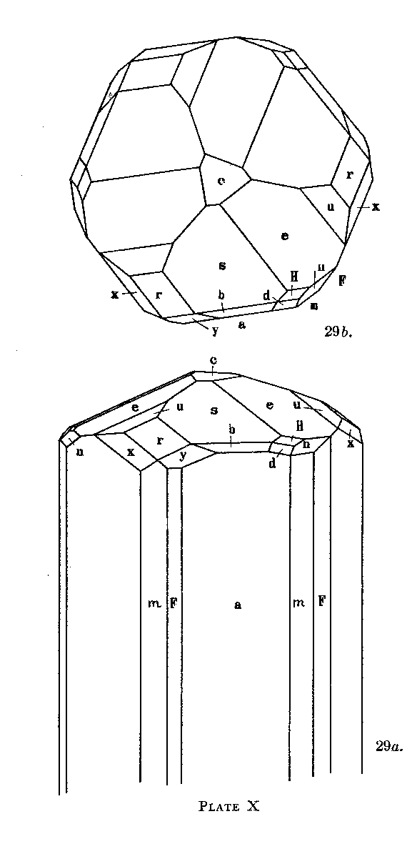
* New forms. Figure 26, Plate IX, shows in part the deeply striated appearance of these crystals. So clear is the material that striations on the back of the crystal are plainly visible through its thickness. Some of them, attached by a prism face, are doubly terminated giving the effect of an enormously elongated rhombohedron. In Plate IX, figure 27a, the elongated rhombohedral aspect due to the oscillation between x and a is represented. As shown on the plan, Plate IX, figure 27b, the upper part of the crystal is bounded by three alternate faces of a. These step down with x and become smaller while the other three faces of a become larger and at the middle of the crystal the six faces of a are in equal development. The same thing occurs on the bottom between the under faces of x and alternate faces of a. Figure 28, Plate IX, shows in plan the distribution of faces as shown on the more highly modified crystals where the rhombohedral aspect is not pronounced. Among the many specimens of crystallized willemite acquired by the Harvard Museum with the Stanton Collection of Franklin minerals was one group distinguished by its extraordinary complexity of crystallization. The prism zone is rounded and vertically striated and the terminal forms are dominantly flat as shown in Plate X, figures 29a and 29b. The crystals are colorless to pale green and show the forms of the following list: c(0001)
r(10 In the following table a summary of the observations made on the new forms is tabulated. The prisms were observed in the striated zone of the stout crystals, and, although they yielded definite signals, they are considered doubtful forms pending confirmation. PLATE X
EXPLANATION OF PLATE X
Figs. 29a and b. Willemite. Clinographic and basal projections of a crystal showing the forms c(0001), a(11
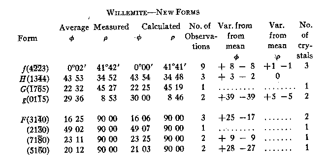
WILLEMITE-NEW FORMS
MISCELLANEOUS. The following minerals have been definitely recognized on Franklin or Sterling Hill specimens and are to be added to the locality list: Anhydrite-Franklin. With these additions the author's list of minerals from these localities now numbers 128 definite species. An explanation for the great abundance and variety of new facts obtained of recent years regarding the Franklin minerals lies chiefly in two circumstances. The active development to deep levels of the Sterling Hill mine has brought to light a host of new data. More important, however, than this is the watchful interest of the chemical staff of the New Jersey Zinc Company. Every new occurrence of minerals in the mines is noted, material secured and analyzed, and its mineralogical nature established. That this is a new state of affairs at Franklin will be apparent to all who have in earlier years been familiar with the local conditions. The author desires to acknowledge his great indebtedness to the New Jersey Zinc Co. for permission to publish the many analyses made by their Franklin chemists. NOTES 1 Palache and Lewis, Am. Mineral., 12, April, 1927. 2 Am. Mineral., 12, 149, 1927. In the figure in this paper showing cahnite, the letter b, wherever it appears should be changed to a. 3 Am. Jour. Sci. , 5, 289, 1898. 4 This number, page 334. 5 This number, page 258. 6 Gordon, 5. G., Proc. Acad. Sci. Phil., 79, 207; 1927. 7 Am. Jour. Sci., 30, 283, 1910. 8 Am. J. Sc., 8. 339, 1899. 9 Am, J. Sc., 29,177.1910. 10 Min. de la France et ses Col., 3, 86, 1901. Figure 56. 11 Bull. de la Soc. Min. de France, Vol. 42, 99, 1919. 12 Proc. Acad. Nat. Sci. Phila., 74, 105, 1922.
|