| Home | AmMin | GMR | RiMG | Collectors Corner | Directory | Short Courses | |
|
|
|||||||

|
|
Volume 59, pages 621-622, 1974
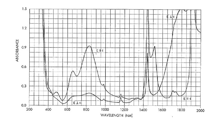
Optical Spectroscopy of Green Vanadium Apophyllite from Poona, India1 GEORGE R. ROSSMAN Department of Geological and Planetary Sciences, California Institute of Technology, Pasadena, California 91109Abstract Emission spectrographic analysis of a light green apophyllite from Poona, India, indicates that 1600 ppm V and 350 ppm Mn are the principal trace constituents. Only 70 ppm Fe was found. Optical spectra have been obtained which indicate that tetravalent vanadium is primarily responsible for the color and dichroism of this material. Instead of being colorless, as is more commonly true in this region, some specimens of apophyllite, KCa 4Si8O20(F,OH)·8H2O, from the Poona region have a pleasing light green color. Because this color resembles that of a variety of silicates containing ferrous iron, it has logically been assumed that these apophyllites were colored by divalent iron. Both optical spectroscopic studies and chemical analyses have demonstrated, however, that this belief is incorrect.The samples examined consisted of transparent green terminated crystals, generally less than one centimeter long, projecting out of a white mass of apophyllite. They are dichroic, ranging from blue-green, E || c, to pale yellow-green, E (perpendicular) c. Their identity was confirmed by infrared spectroscopy which indicated that the spectrum of the green material is identical to that of colorless apophyllite from Poona which in turn is identical to the portion of the Michigan apophyllite spectrum reported by Colville and Anderson (1971). An emission spectrographic analysis indicated that vanadium is the most abundant trace transition metal (Table 1). A report of the analysis of a colorless apophyllite from Poona (Belsare, 1969) did not report the presence of vanadium. The single crystal studied optically was free of internal defects, had nearly flat, parallel crystal faces, and was uniaxial negative in the center but graded to biaxial negative near the edges (with 2 V up to 12°). Optical absorption spectra (Fig. 1) show that prominent absorption bands centered at 492, 646, and 829 nm are responsible for the green color in the E || c spectrum. In the near-infrared the prominent features are at 1442 nm and 1517 nm polarized parallel to c, about 1795 nm polarized perpendicular to c, and the intense feature at about 1921 nm whose polarization was not determined. The features at 1442 and 1517 nm are, respectively, the first overtones of the OH stretching motion which occurs at 3556 cm -1 in the infrared spectral region, and a stretching motion of the water molecules in the 3000-3400 cm-1 region.The absorption bands at 492, 646, and 829 nm responsible for the color are attributable to vanadium and manganese. Two oxidation states of vanadiumV 3+, and V4+ as the VO2+ ion-would be considered reasonably likely to be found in the apophyllite. In an electron spin resonance study of apophyllite from Nidy, Siberia, Marfunin, Bershov, and Mineeva (1966) obtained the signal of VO2+. The VO2+ was believed to be present in both the eight-fold tetragonal-prismatic potassium site and in the seven-fold capped trigonal-prismatic calcium site. The optical results indicate that the bulk of the vanadium is present as the VO2+ ion. The apophyllite spectra resemble the spectra of crystalline VOSO4·5H2O and vanadyl acetylacetonate solutions (Ballhausen and Gray, 1962; Bernal and Rieger, 1963). In the spectrum of crystalline vanadyl sulfate bands at 13,100 and 16,000 cm-1 polarized perpendicular to the V=O bond axis were assigned to the 2B2 --> 2E and 2B2 --> 2B1 transitions. The corresponding transitions in apophyllite are assigned to the 12,000 cm-1 (829 nm) and 15,500 cm-1 (646 nm) bands respectively. Although the band at 20,400 cm-1 (492 nm) could be analogous to the 25,300 cm-1 band in the vanadyl acetylacetonate spectrum, it appears more appropriate to attribute it to Mn3+ in view of the fact that minerals containing Mn3+ in relatively symmetrical environments have prominent absorptions in the range 480-560 nm. If all Mn is assumed to be present in the trivalent state, ε in the E || c polarization is 27 l.mole-1 cm-1.
|
|
FIG. 1. Optical absorption spectrum of apophyllite from Poona, India. The top
curve is for incident light polarized E || c; bottom curve, E (perpendicular)
c. Sample: 2.64
mm thick self-supporting crystal. 296°K spectrum. Absorbance in units of log I0/I.
TABLE 1. Emission Spectrographic Data
The optical data do not provide a clear indication that the vanadium is distributed between the calcium and potassium sites. To the contrary, the close similarity between the VOSO 45H2O spectrum and the apophyllite suggest that if the vanadium is a true substitutional impurity, then rather than conforming to the seven- or eight-coordinate site of the ion which it replaced, the site will be modified to conform to the energetics of the vanadium in a manner similar to that reported by Ghose and Tsang (1973) where iron substituting for aluminum in a variety of minerals apparently distorts the site to new geometry more favorable for the iron.Acknowledgment The author wishes to thank R. H. Currier for providing the specimens used in this study. References BALLHAUSEN, C. J., AND H. B. GRAY (1962) The electronic structure of the vanadyl ion. Inorg. Chem. 1, 111-122. BELSARE, M. R. (1969) A chemical study of apophyllite from Poona. Mineral. Mag. 37, 288-289. BERNAL, I., AND P. H. RIEGER (1963) Solvent effects on the optical and electron spin resonance spectra of vanadyl acetylacetone. Inorg. Chem. 2, 256-261. COLVILLE, A. A., AND C. P. ANDERSON (1971) Refinement of the crystal structure of apophyllite I. X-ray diffraction and physical properties. Am. Mineral. 56, 1222-1233. GHOSE, S., AND T. TSANG (1973) Structural dependence of quadrupole coupling constant e 2qQ/h for 27Al and crystal field parameter D for Fe3+ in aluminosilicates. Am. Mineral. 58, 748-755.MARFUNIN, A. S., L. V. BERSHOV, AND R. M. MINEEVA (1966) La résonance paramagnetique électronique de l'ion VO 2+ dans le sphène et l'apophyllite et de l'ion Mn2+ dans la trémolite, l'apophyllite et la scheelite. Bull. Soc. Franc. Mineral. Cristallogr. 89, 177-183.NOTES 1 Contribution No. 2397, Division of Geological and Planetary Sciences, California Institute of Technology. Manuscript received, December 27, 1973; accepted for publication, January 30, 1974. |
||||||||||||||||||||||||||||||||||||||||