 |
|
|
Topology of syntectonic melt flow networks in the deep crust: inferences from three-dimensional images of leucosome geometry in migmatites Mary Anne Brown1, Michael Brown1*, William D. Carlson2, and Cambria Denison2 1 Laboratory for Crustal Petrology, Department of Geology University of Maryland, College Park, MD 20742, USA2 Department of Geological Sciences, University of Texas at Austin, Austin, TX 78712, U.S.A*E-mail: mbrown@geol.umd.edu
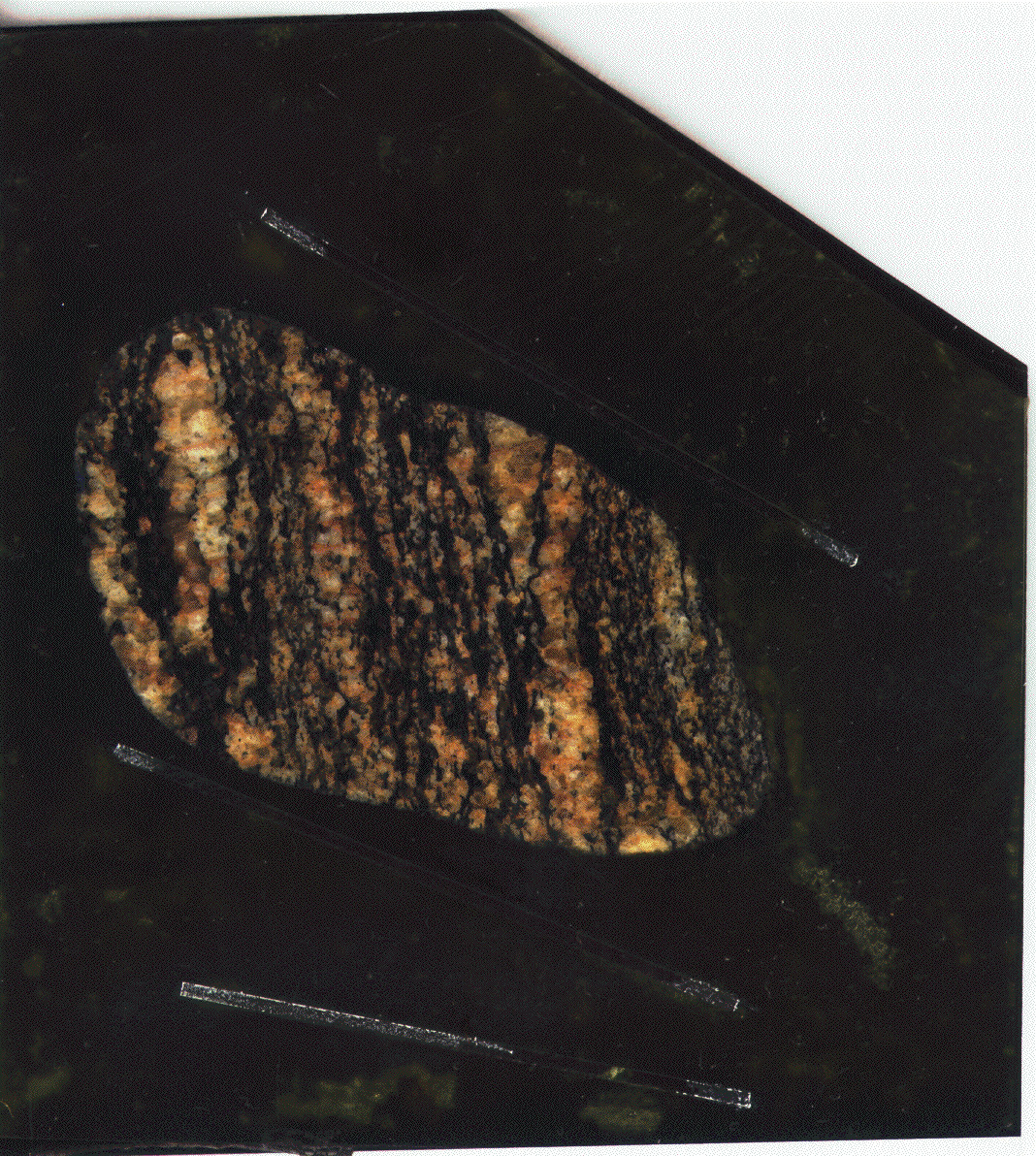
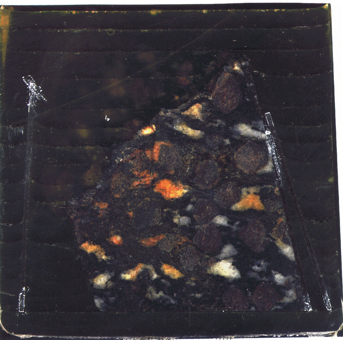
Web-Based Figures Fig. 2. (a; 968 Kb) Full-size high-resolution color image of the stromatic migmatite. The image was created by optical scanning of the first slice of a serially ground block of epoxy in which the sample was embedded (upper center of field of view). The white linear features in the epoxy (lower center of field of view) are triangular glass slides used to measure slice thickness. (b; 1.96 Mb) Animated gif of contrast-enhanced gray scale representations of 39 serially ground slices of the stromatic migmatite. First and last slices are outlined in white. Animation cycles from first to last slice. Fig. 3. (a; 1.08 Mb) Full-size high-resolution color image of the migmatitic garnet-amphibolite. The image was created by optical scanning of the first slice of a serially ground block of epoxy in which the sample was embedded (center of field of view). The white linear features in the epoxy (right of field of view) are triangular glass slides used to measure slice thickness. (b; 1.50 Mb) Animated gif of contrast-enhanced gray scale representations of 42 serially ground slices of the migmatitic garnet-amphibolite. First and last slices are outlined in white. Animation cycles from first to last slice. Fig. 4. (c; 3.61 Mb) Animated gif of gray scale inverted images of 108 HR X-ray CT scans of the stromatic migmatite. First and last slices are outlined in white. Animation cycles from first to last slice. Fig. 5. (c; 10.9 Mb) Animated gif of gray scale inverted images of 112 HR X-ray CT scans of the magmatitic garnet amphibolite. First and last slices are outlined in white. Animation cycles from first to last slice. Fig. 7. (e; 3.57 Mb) Animated gif of three-dimensional projection of the stromatic migmatite rotating through 360o. This projection was created by compiling the two-dimensional representations created by serial grinding into a stack using NIH Image. Each slice in the stack can be thought of as a one-pixel-thick plane separated by the thickness of material cut off the sample (~1.5 mm). The stack was rotated around a virtual horizontal axis in the plane of the page. A scale bar is not presented because this is a three-dimensional projection; however, Fig. 2a contains the scale for the first representation in the stack. The light-colored parts of the migmatite (leucosome and felsic minerals in the mesosome) are white and gray. The dark parts of the migmatite (melanosome and mafic minerals in the mesosome) are transparent. Because the melanosome is transparent, this projection presents the three-dimensional structure of individual planar leucosomes. Fig. 8. (e; 787 Kb) Animated gif of three-dimensional projection of the migmatitic garnet-amphibolite rotating through 360o. This projection was created by compiling the two-dimensional representations created by serial grinding into a stack using NIH Image. Each slice in the stack can be thought of as a one-pixel-thick plane separated by the thickness of material cut off the sample (~1.5 mm). The stack was rotated around a virtual horizontal axis in the plane of the page. A scale bar is not presented because this is a three-dimensional projection; however, Fig. 3a contains the scale for the first representation in the stack. The light-colored parts of the migmatite (leucosome) are white and gray. The dark parts of the migmatite (melanosome and garnet) are transparent. Because the melanosome is transparent, this projection presents the three-dimensional structure of the leucosome. Fig. 9. (e; 6.47 Mb) Animated gif of three-dimensional projection of the stromatic migmatite rotating through 360o. This projection was created by compiling the two-dimensional representations created by HR X-ray CT into an animation created using VoxBlast®. The three-dimensional image produced is superior to the one created using NIH Image using two-dimensional representations created by scanning after serial grinding (Fig. 7) because the HR X-ray CT scans are more closely spaced, and VoxBlast® calculates a virtual volume from the stack of two-dimensional representations. The sample rotates in the animation around a virtual horizontal axis in the plane of the page. A scale bar is not presented because this is a three dimensional projection; however, Fig. 4a contains the scale for the first representation in the stack. The less-dense parts of the migmatite (leucosome and felsic minerals in the mesosome) are white and gray. The denser parts of the migmatite (melanosome and mafic minerals in the mesosome) are transparent. Because the melanosome is transparent, this projection presents the three-dimensional structure of individual planar leucosomes. Fig. 10. (e; 8.23 Mb) Animated gif of three-dimensional projection of the migmatitic garnet-amphibolite rotating through 360o. This projection was created by compiling the two-dimensional representations created by the HR X-ray CT into an animation created using VoxBlast®. The three-dimensional image produced is superior to the one created using NIH Image using two-dimensional representations created by scanning after serial grinding (Fig. 8) because the HR X-ray CT scans are more closely spaced, and VoxBlast® calculates a virtual volume from the stack of two-dimensional representations. The sample rotates in the animation around a virtual horizontal axis in the plane of the page. A scale bar is not presented because this is a three dimensional projection; however, Fig. 5a contains the scale for the first representation in the stack. The less-dense parts of the migmatite (leucosome) are white and gray. The denser parts of the migmatite (melanosome and garnet) are transparent. Because the melanosome is transparent, this projection presents the three-dimensional structure of the leucosome. Fig. 14. (g; 538 Kb) Animated gif of a three-dimensional projection of a single connectivity "tree" in the migmatitic garnet-amphibolite. The stack rotates through 360o around a virtual vertical axis in the plane of the page. (Note that this axis of rotation is different from the axis in Figs. 7-10). A scale bar is not presented because this is a three-dimensional projection; however, Fig. 5a contains the scale for the first representation in the stack. Note that a complex tree of leucosome throughout the stack is connected to a single leucosome on top of the stack. Fig. 15. (g; 230 Kb) Animated gif of a three-dimensional projection of a single path within the connectivity "tree" in Fig. 14. The stack rotates through 360o around a virtual vertical axis in the plane of the page. (Note that this axis of rotation is different from the axis in Figs. 7-10). A scale bar is not presented because this is a three-dimensional projection; however, Fig. 5a contains the scale for the first representation in the stack. Note that the path includes a few very narrow necks through which fluid must flow. Fig. 16. (e; 15.9 Mb) Animation of projection of false-color three-dimensional image of stromatic migmatite, derived from the stack of two-dimensional representations of the HR X-ray CT scans, created using VoxBlast®. A scale bar is not presented because these are projections of a three-dimensional image. (Fig. 4b contains the scale for the first image in the stack.) The leucosome is rendered transparent in this image. The color of the solid part of the image is brighter for material with higher mineral density, with garnet appearing yellow. Fig. 17. (e; 7.21 Mb) Animation of projection of false-color three-dimensional image of leucosome in the magmatitic garnet-amphibolite, derived from the two-dimensional representations of the HR X-ray CT scans, created using VoxBlast®. A scale bar is not presented because these are projections of a three-dimensional image. (Fig. 5b contains the scale for the first image in the stack.) Garnet associated with the leucosome and the matrix have been rendered transparent in this image. |