| Home | AmMin | GMR | RiMG | Collectors Corner | Directory | Short Courses | |
|
|
|||||||

|
|
Volume 64, pages 151-255, 1979
Fibrous intergrowths of cross muscovite and cross chlorite from shear zones of Pennsylvanian carbonaceous rocks in Rhode Island MARTIN S. RUTSTEIN Department of Geological Sciences Abstract Veinlets of light green to white, fine acicular intergrowths of muscovite and chlorite occur in shear zones of chlorite-grade Pennsylvanian carbonaceous strata from the Portsmouth, R.I., area. Alleghanyan deformation produced a set of shear surfaces containing these intergrowths, oriented with {001} at a high angle to the bounding shear surface. Sander (1930) describes such structures as cross micas. X-ray analysis indicates no interstratification. The intergrowths are well-crystallized muscovite (probably 2M,) and chlorite (trioctahedral and Mg: Fe ~= 2: 1). The phases are not readily distinguishable by light microscopy. However, heat treatment at 825°C destroys the chlorite structure and produces a brown, amorphous, expanded layer which accentuates the intergrowth relationship. Typically, discrete elongate euhedral fibers of muscovite and chlorite are intergrown parallel to [100] or [010]. Also, shorter and euhedral to subhedral fibers of chlorite are commonly observed as inclusions in muscovite. The unusual elongation is believed due to crystal growth into an opening shear zone. Oriented seed crystals in the phyllite matrix initiated growth mainly along the a axis. Subsequent interference with neighboring crystals limited growth in the c and one other direction. Cross fibers developed through relatively unimpeded growth at the tips of crystals. Introduction In 1976, near Portsmouth, R.I., I observed boulders of carbonaceous phyllite containing cross-veinlets of a material with pronounced fibrous properties. The boulders are derived from carbonaceous beds of the Pennsylvanian Rhode Island Formation, which was metamorphosed to chlorite grade during the Alleghanyan metamorphic event. Presumably, the veinlets formed during the metamorphic event. Casual inspection suggested the veinlet material might be chrysotile asbestos. However, closer field testing of the physical properties of the material (crushing and rubbing between fingertips) showed that the material did not mat together as is typical of asbestos. Instead, the fibers were slippery and felt almost talc-like in their softness. Moreover, syngenetic asbestos has never been reported from chlorite-grade carbonaceous phyllites. If the material were asbestos, the particular paragenesis would be notable. The literature on the area mentions the occurrence of fibrous quartz in these strata. This suggestion may arise from the work of Quinn and Glass (1958) who studied these units and reported the presence of veinlets of "quartz, calcite, pyrite and aphrosiderite (partially replaced by fibrous quartz)," aphrosiderite being a varietal name for ripidolite. However, the bulk of the fibrous material observed by the writer is too soft to be quartz. The material was subsequently X-rayed and identified as a fibrous intergrowth of two typically platy minerals-muscovite and chlorite. This paper reports on the morphologic and intergrowth relations of the phases and suggests a mechanism for the origin of the unusual habit. Techniques The larger (to 5mm long) fibers were easily handpicked from the matrix and separated for routine petrographic, SEM, and X-ray study. Smaller fibers (a few mm long) were also studied but were handpicked using a binocular microscope. Other than length, there are no physical differences between the two fractions.
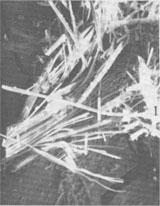
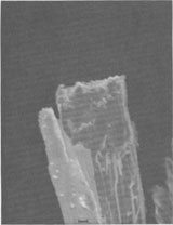
For X-ray identification, a General Electric XRD7 unit employing Cu radiation and a Ni filter was employed. Medium slit systems (3°,MR, and 0.1°) were used for initial studies and fine slits (1°,MR, and 0.1°) for the remaining work. Unoriented smear slides for diffraction analysis were prepared by cementing (with Duco cement dissolved in acetone) finely ground sample and sample plus ground silica glass onto a petrographic glass slide. Because the former produced excellent basal reflections, no serious attempt was made to produce oriented slides by the several standardized methods. Heating of the fibers was done in a muffle furnace. Samples were ground under acetone, placed in a silica glass dish and heated at 425° for 30 minutes, 700° for 20 minutes, and 825°C for 15 minutes. The material was also characterized using an AMR 1200 Scanning Electron Microscope and PGT non-dispersive energy analyzer. Results The X-ray diffraction data indicate two well-crystallized, discrete phases. The reflections are integral basal reflections of muscovite and chlorite. There are no diffuse or intermediate reflections indicating a random or non-random interstratified mix of sheet silicates. Heated and rapidly cooled samples were also X-rayed. Those heated to 325°C show no change. Those heated to 700°C display an intensification of the chlorite (001) reflection and no change in the muscovite reflections. The samples heated to 825°C reveal no fundamental changes in muscovite intensities or d values, but do indicate slight reflection broadening. Chlorite, however, is now absent, having been converted to what appears to be an amorphous or highly disordered residue. Overall, the sharpness of the reflections, their d values, and their retention to various temperatures indicate that the material consists of chlorite and muscovite. Analysis of the individual and collective reflections of chlorite as suggested by Grim (1968) and Carroll (1969) indicates that the chlorite is a trioctahedral Mg-rich chlorite, but contains more than a negligible amount of iron. Such evidence includes: (a) odd hkl reflections being stronger than even hkl reflections; (b) the presence of even hkl reflections; (c) heating aliquots to 700°C intensifying the (001) reflection and diminishing the others; (d) the intensity ratio (I002 + I004)/I003 (Petruk, 1964) indicating that of the six octahedral positions, about four are filled with Fe, and the remainder with Mg and Al. Other supporting evidence includes: (a) qualitative energy-dispersal analyses indicating slightly more Fe than Mg; and (b) the change of' chlorite heat-treated at 825°C to a brownish breakdown product interpreted as an iron-oxide colored silicate. The muscovite was determined to be well-crystallized, ordered, and probably the 2M1 polymorph. In similar samples Quinn and Glass (1958) report coexisting muscovite and paragonite; however, no evidence of paragonite was found in this study. Moreover, the Na content of the muscovite is barely detectable by qualitative energy-dispersive analysis. The matrix material was also X-rayed. Although complex, the pattern indicates coexisting chlorite, muscovite, quartz, and graphite. Optical examination does not readily suggest the existence of two discrete phases. The fibers occur as light green to white, fine acicular crystals oriented at a high angle to the bounding shear surfaces. Luster and coloration are generally constant parallel to fiber length, but take on a somewhat chatoyant effect at right angles to the direction of elongation. With oil-immersion techniques, the material appears to be either (a) relatively homogenous; (b) twinned with the twin plane parallel to fiber length; (c) composed of two discrete phases with very similar indices of refraction; or (d) composed of two similar phases in varying orientations with respect to fiber length. SEM examination at lower magnifications reinforces the petrographic study. Figure 1 shows the fibrous properties of the mixture and indicates the cleavage. Figure 2 is a SEM photomicrograph of a fiber tip. The mass shows several cleavages which would form a tabular crystal were it not for the existence of elongation beyond the field of view. The fibrous properties appear to be one of scale - normal platy development on a microscopic scale, but an unusual elongation in one crystallographic direction. The point will be considered below. The existence of two phases in Figure 2 is suggested by the differential deposition of copper. Energy-dispersive line-scan analysis clearly reveals the existence of the two phases. Optical examination of the material heated to 825°C also shows the coexistence of the two phases indicated by the X-ray analysis. At the high temperature, the chlorite altered to an expanded, brown, lathlike "phase" which alternates with a transparent phase. Typically, altered "chlorite" is included in muscovite. The reverse relationship is less common. The expanded, brown material is typically curved or bent and exhibits cracks (presumably tension) resulting from the cooling of an expanded lattice. The general intergrowth pattern is chlorite and muscovite fibers packed parallel to fiber length, with lengths continuous or discontinuous with respect to the bounding shear surfaces. With this information, reexamination of unheated material enabled recognition of the slight differences between the phases. The apparently homogenous nature of the mixture is due to the extremely narrow fiber width (usually < 0.005mm); similar indices of refraction (n ~= 1.57); and similar color (light green to colorless). Extinction angle varies across a fiber packet. Fibers of muscovite were typically length-fast, but length-slow grains were not uncommon. Length-fast indicates an orientation with microscope view normal or nearly normal to (001). Conversely, length-slow indicates a grain oriented parallel to the cleavage trace (e.g., Kerr, 1959, p. 386). These orientations indicate that fiber length is parallel to a or b. No example of fiber composed only of chlorite was observed. However, although the chlorite is typically very fine-grained and included in more massive muscovite, a few grains are sufficiently large for limited optical study. The few grains noted are length-slow, indicating that fiber length is parallel to (001) (e.g., Kerr, 1959, p. 396). Three other observations indicate that a or b and not c is the elongate direction: (a) petrographic and SEM examination (see Fig. 2) of fiber tips show a ragged surface instead of cleavage; (b) SEM examination (see Fig. 1, 2) of fiber bundles reveals cleavage parallel to the long axes of the fibers; and (c) the strong basal reflections of semi-oriented materials are consistent with {001} parallel to the long axes of the majority of grains. One piece of evidence indicates a rather than b as the elongate direction. Heating the sample results occasionally in freeing from the matrix masses of muscovite large enough to provide an optical interference figure. The few grains so studied generated an obtuse bisectrix figure, indicating a microscope view along the Z optical axis (or b crystallographic direction). Cleavage was parallel to fiber length. Hence fiber length must correspond with a.
From the evidence, I conclude that the fibers exist as aggregates of muscovite and chlorite. Fiber length is commonly parallel to a or b rather than c. It is possible, but unproven, that the a elongation is predominant. Discussion The material found in the shear fractures is a fibrous intergrowth of discrete and homogenous masses of muscovite and chlorite, two minerals which usually occur as platy aggregates. Hence, it is of interest to consider what factors could cause the unusual morphology. For minerals which do not have structures which promote fibrous growth (e.g., gypsum, var. satin spar), a common explanation for such fibrous development is that constraints on morphology develop from the nature of the bounding surfaces, e.g. in a "container" for the growing crystals (Correns, 1969, p. 164-165; Grigor'yev, 1965, p. 190-191). After the nucleation stage, embryonic crystals grow and develop in a tabular, confined space. Impingement of growing crystals and competition for solute lead to the elimination of many crystallites, and the few survivors have shapes determined by the "container" in which they grew. The mechanism develops an aggregate of parallel, columnar crystals. The veinlets of muscovite and chlorite occur in shear fractures developed during deformation. For involvement in a deformation cycle, the environment of crystal growth and subsequent deformation can be modeled as a stress field or as a non-stress field. In the former case, micas commonly undergo mimetic crystallization (Turner and Weiss, 1963, p. 440-442) and develop with (001) normal to applied stress. This results in the commonly observed schistosity of micaceous rocks. As an intermediate type of stress situation, Grigor'yev (1965, p. 199-200) refers to an example of crystal growth in a slowly opening fissure. Tectonic control is not completely absent but is a minor parameter in controlling morphology. In his model, many possible orientations of crystals develop in the fissure. Individual crystals become elongated as the crystal grows into the opening fissure or vein. However, the random orientations contrast with the regular, parallel orientations reported here. In a non-stress field, micas can develop at least two extremes of morphology. In a pegmatite (crystallizing in a hydrostatic environment) muscovite crystals often develop crystals elongate to c, but with marked development of a and b. These blocky, columnar masses are in contrast to the fibers reported here, which have substantial growth along only one axis (a or b) and relatively negligible growth along the other two axes. In contrast, Sander (1930, p. 208, 212-213) identifies a morphological class of micas which has {001} normal to the shear surfaces bounding the crystals. Referring to these as "cross micas," he suggests they are the result of post-tectonic crystallization events occurring in tectonically opened fractures. Why they grew with this particular orientation is not understood. The orientation of cross micas described by Sander is similar to the fibers reported here. Any model explaining the specific occurrence, and perhaps also a more general explanation, must result in crystals elongate along only a or b, with the two remaining axial lengths being relatively negligible. A proposed model suggests how the development of muscovite and chlorite might be affected by the tectonic forces operating on a volume of rock containing a growing crystal. During chlorite-grade metamorphism, the host rock should still be relatively "wet" with fluids. These fluids will probably be close to or in actual equilibrium with chlorite and muscovite in the host-rock matrix and, for a given pressure and temperature, can be considered to be saturated with respect to muscovite and chlorite. If a shear surface in these semiporous phyllites begins to open, fluids will diffuse and migrate toward the opening. Upon reaching the solid-void interface, they will quickly become supersaturated due to the drop in pressure. However, from Grigor'yev's model, nucleation in the non-stress field of an open fracture should produce a random and more variable set of orientations than those observed here. Still, a means of controlling the orientations of growing crystals exists as a result of the forces which produce the shearing. I suggest that the shearing forces opening the rock also affect those matrix detrital muscovite (and chlorite) flakes which are near the fracture surface. The forces opening the fracture can induce slight rotation, bending, and/or recrystallization. Then, any nearby slightly elongate mineral exposed at the interface of rock-void will become oriented at a slightly higher angle to the shear fracture. The elementary but often workable Law of Bravais can be applied to estimate the original geometry of the matrix grains. The predicted growth rates for muscovite are about 4:2:1 for (100):(010):(001). The bonding factor should enhance the growth of (100) and (010) relative to (001) (Dowty, 1976). For chlorite, the differences are less marked, the ratios being 3:1.5: 1. Thus, the platy matrix grains should be slightly axially elongate in the decreasing order a, b, and c. Grigor'yev (1965, p. 198-203) suggests that once crystal growth begins in an opening fissure, continued opening will enhance the existing morphology. The present model suggests that initially the matrix crystals are slightly elongate. Subsequently, they are disturbed by the shearing stresses and act as seed crystals in the presence of supersaturated fluid. The existing morphology will be enhanced along the direction of inherited growth and the opening shear zone (the container). The fastest growth will occur at the tips of crystals (presumably on {100} or {010}). This growth probably occurs in a relatively unimpeded and stress-free environment, since accumulation in any other direction will be limited by the structure of the phases, by fissure walls, or by neighboring crystals. Large numbers of neighbors are probable, given the number of seed crystals capable of initiating growth in the presence of the pervasive supersaturated fluid. As a result of interference with adjacent crystals, the tabular geometry with (100):(101):(001) being about 4:2:1 is overwhelmed by growth at fiber tip (recall Fig. 2). Thus, the geometry approximates ∞ : 2: 1 (or possibly (4: ∞ : 1)), and the resulting crystalline aggregate is a mass of fibrous muscovite and chlorite. Acknowledgments The writer thanks Drs. J. Skehan and D. Murray for their leadership of the 1976 NEIGC field trip on which the samples reported here were discovered. References Carroll, D. (1969) Clay Minerals: A Guide to their X-Ray Identification. Geol. Soc. Am. Spec. Pap. 126. Correns, C. W. (1969) Introduction to Mineralogy, 2nd ed. (trans. W. D. Johns). Springer-Verlag, New York. Dowty, E. (1976) Crystal structure and crystal growth: I. The influence of internal structure on morphology. Am. Mineral., 61, 448-459. Grigor'yev, D. P. (1969) Ontogeny of Minerals, [trans. Israel Prog. for Scientific Trans. Staff.] Daniel Davey, New York. Grim, R. E. (1968) Clay Mineralogy, 2nd ed. McGraw-Hill, New York. Kerr, P. F. (1959) Optical Mineralogy, 3rd ed. McGraw-Hill, New York. Petruk, W. (1964) Determination of the heavy atom content in chlorite by means of the X-ray diffractometer. Am. Mineral., 49, 61-71. Quinn, A. W. and H. D. Glass (1958) Rank of coal and metamorphic grade of rocks in the Narragansett basin of Rhode Island. Econ. Geol., 53, 563-576. Turner, F. J. and L. E. Weiss (1963) Structural Analysis of Metamorphic Tectonites. McGraw-Hill, New York.
Manuscript received, December 13, 1977; |