| Home | AmMin | GMR | RiMG | Collectors Corner | Directory | Short Courses | |
|
|
|||||||

|
|
Volume 64, pages 169-171, 1979
Authigenic mitridatite from the Shungura Formation, southwestern Ethiopia ROBERT J. ROGERS AND FRANCIS H. BROWN
Department of Geology and Geophysics, University of Utah Abstract Mitridatite from lacustrine sediments in the Shungura Formation of southwestern Ethiopia occurs in small crystals (<2 µm) with hydroxy-apatite and carbonate-apatite. An approximate structural formula for the mitridatite is Ca6(H2O)6[Fe3+8.2Mn3+0.8O6(PO4)9]·3H2O, placing the mineral in the mitridatite-robertsite series. Mineral associations in the lacustrine sediments suggest that the mitridatite coprecipitated with hydroxy-apatite, following partial dissolution of carbonate-apatite fish scales and bones. Introduction Mitridatite was first described in the early part of this century from oolitic sedimentary iron ores of the Kerch and Taman Peninsulas, U.S.S.R., but remained an ill-defined species due to the lack of coarse-grained material for study. Tarnovskiy and Kashayeva (1969) and Moore (1964, 1974) have reported mitridatite from pegmatites. Moore (1974), working with coarser material than was previously available, confirmed the specific status of mitridatite and suggested equivalence between the pegmatitic material and material from Russian sedimentary sources, which had been studied most recently by Chukhrov et al. (1958). In the same study Moore also described a newly discovered manganese phosphate, robertsite, and showed it to be isotypic with mitridatite. He also suggested that the two minerals form a series. In this note we report the occurrence of a member of that series, a Mn-bearing mitridatite, in late Cenozoic lacustrine sediments in Ethiopia, where it has apparently coprecipitated with hydroxyapatite. Occurrence The mitridatite was collected in 1974 during fieldwork of the Omo Research Expedition. It occurs in the Shungura Formation of southwestern Ethiopia. For details of the stratigraphy of this formation, the reader is referred to de Heinzelin et al. (1976); only a brief summary is presented below. The Shungura Formation is of Pliocene/Pleistocene age, and has been divided into twelve members on the basis of major intercalated tuffs lettered A through L from bottom to top, with a Basal Member defined as the sediments below Tuff A. Each member is made up of its basal tuff and sediments above it up to the next major tuff. The members have been divided into submembers on the basis of internal changes in sediment type, and these are labelled numerically from the base of each member. The sediments from the Basal Member through submember G-13 reflect deposition in a fluvial environment for the most part, although minor lacustrine incursions occur. A major lacustrine sequence begins in submember G-14 and continues to G-26. This sequence is followed by fluvially deposited sediments which extend to the top of the formation, although again minor lacustrine incursions are recorded. Dessication cracks are preserved at many levels, indicating that at least this part of the lake was shallow, and that the sediments were subaerially exposed at various times during their deposition. Gypsum and halite appear in many of the clays, indicating that the lake was relatively saline. Calcite nodules, concretions, and algal mats are present sporadically, indicating that the lake was probably alkaline, but the salinity was not so high that ostracods, gastropods, and fish found it intolerable. Mitridatite was first identified from submember G-22 by X-ray diffraction methods during clay mineralogy studies. Further X-ray work on material of similar appearance revealed the presence of mitridatite in samples from G-25 and the contact of G-24 and G-25. In all instances the mitridatite is closely associated with fish scales and/or bones in silty clay or clay. The mitridatite is very fine-grained, and X-ray diffraction work and scanning electron microscopy show it to be admixed with hydroxy-apatite, carbonate-apatite (fish skeletal remains), quartz, smectite clays, and hydrous iron oxides of undetermined species.
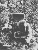
Description Mitridatite occurs in submember G-22 as impure, earthy, olive-green blotches and masses. In the other submembers the mitridatite, again impure, occurs in layers less than 3 mm thick, concentrically surrounding fish bones and scales in concretionary features from 1.5 to 10 cm in diameter. The layers were observed both immediately surrounding the fish remains, and separated from the remains by as much as 1 cm. These layers are darker green than the earthy masses of G-22, probably due to a higher mitridatite content. Mitridatite was never found in pure masses of sufficient size to be isolated for detailed X-ray and chemical studies. Examination of mitridatite-rich sediments from submember G-22 with a scanning electron microscope revealed small porous areas in the sediment lined with very small, composite mitridatite crystals. These composite crystals, saddle-shaped and feathery in appearance, average between 2.0 and 2.5 µm in their greatest dimension. They form aggregates and are generally associated with prismatic crystals of hydroxy-apatite up to about 15 µm long. The hydroxy-apatite crystals appear both on the mitridatite, and with mitridatite on them (Fig. 1). Mitridatite crystals were not observed in samples from the concretionary features, due to the more compact nature of the material.The mitridatite was examined in immersion oils with a petrographic microscope, but because of the extremely fine-grained nature of the material and the composite nature of the crystals, little information could be obtained. The color ranges from greenish-brown to yellowish-green, which agrees with the colors reported by Moore (1974), but actual pleochroism, reported by Moore, was not observed, probably because of the small grain size. The material has a mean index of refraction between 1.770 and 1.785, varying from sample to sample possibly because of varied degrees of hydration. X-ray data In all X-ray diffraction patterns the mitridatite was found to be admixed with hydroxy-apatite and/or carbonate-apatite, which have lines which coincide with or are very close to mitridatite lines. In all but a few cases the apatite and mitridatite lines could be resolved with the diffractometer by using slow scanning speeds. However, because of the admixed apatite, intensities of some mitridatite lines could not be obtained from the films. The X-ray d-spacing data are in good agreement with those of Moore (1974) for mitridatite. Chemistry Semiquantitative chemical information was obtained for the mitridatite with an energy-dispersive X-ray spectrometer coupled to a scanning electron microscope. The mitridatite was found to contain manganese in more than trace amounts. The presence of manganese places the mitridatite in the mitridatite-robertsite series, first suggested by Moore (1974). A pure sample of the mitridatite could not be isolated for chemical analysis. However, a sample of mitridatite-rich material from G-22 was dissolved and analyzed for iron and manganese by atomic absorption spectrometry. The same sample was X-rayed and found to contain only mitridatite and hydroxy-apatite in detectable amounts. Further, no iron or manganese were detected in the apatite with the energy-dispersive spectrometer. Therefore all the iron and manganese were assumed to reside in the mitridatite. Using the structural formula proposed by Moore and Araki (1977) and assuming all the iron to be ferric, the following formula was calculated: Ca6(H2O)6[Fe 3+8.2Mn3+0.8O6(PO4)9]·3H2O. It should be emphasized that this is offered only as an approximate formula for the material. Because of small amounts of hydrous iron oxides observed under the petrographic microscope in some of the mitridatite samples, the above formula is believed to represent the upper limit of the Fe: Mn ratio. The semiquantitative energy-dispersive X-ray data support this conclusion.Paragenesis Mitridatite crystals such as those in Figure 1 and their occurrence in concretionary features leave no doubt that the mineral is authigenic. Further, the material studied was always found in close association with fish scales and/or bones. X-ray diffraction patterns of several of these bones were found to match carbonate-apatite. The partial dissolution of carbonate-apatite fish remains served as the source of phosphate for the mitridatite and hydroxy-apatite which precipitated nearby. The hydroxy-apatite was observed in the form of well-developed crystals in intimate association with the mitridatite. The spatial relationship between the mitridatite and the hydroxyapatite suggests that the two minerals were coprecipitated.
Conclusion The mitridatite described here represents a positively identified member of the mitridatite-robertsite series, which appears to have formed by direct precipitation from solution. In all previously reported occurrences mitridatite was observed to have formed from the alteration of transition-metal phosphates. Mitridatite should be suspected in the future as a possible authigenic phase in sediments containing vertebrate remains. Acknowledgments Field work of the Omo Research Expedition was supported by grants from the Wenner-Gren Foundation, the National Geographic Society, and the L. S. B. Leakey Foundation in 1974, and laboratory work was supported by a grant from the University of Utah Research Committee. This support is gratefully acknowledged. Dr. Adolf Pabst kindly read the manuscript in preliminary form and made many corrections and useful criticisms for which we thank him. References Chukhrov, F. V., V. A. Moleva and L. P. Ermilova (1958) New data on mitridatite. Akad. Nauk. S.S.S.R. Izuestiya, Ser. Geol., No. 8, 16-26 (translated from Russian). de Heinzelin, J., P. Haesaerts and F. C. Howell (1976) Plio-Pleistocene formations of the Lower Omo Basin with particular reference to the Shungura Formation. In Y. Coppens, F. C. Howell, G. Ll. Isaac, and R. E. F. Leakey, Eds., Earliest Man and Environments in the Lake Rudolf Basin, p. 24-49. University of Chicago Press, Chicago. Moore, P. B. (1964) Notes on some Black Hills phosphates. Am. Mineral., 49, 1119-1122. ________ (1974) 1. Jahnsite, segelerite and robertsite, three new transition metal phosphate species. 11. Redefinition of overite, an isotype of segelerite. III. Isotypy of robertsite, mitridatite, and arseniosiderite. Am. Mineral., 59, 48-59. _________ and T. Araki (1977) Mitridatite, Ca 6(H2O)6[FeIII9 O6(PO4)9]·3H2O. A noteworthy octahedral sheet structure. Inorg. Chem., 16, 1096-1106.Tarnovskiy, G. H. and G. M. Kashayeva (1969) Mitridatite found for the first time in pegmatite in the USSR. Dokl. Acad. Sci. U.S.S.R., Earth Sci. Sect., 183, 153-155 (translated from Russian).
Manuscript received, April 13, 1978;
|